New imaging techniques allow scientists to map sugar structures on cell surfaces with unprecedented detail, revealing cellular states previously hidden. This development, detailed in recent publications, marks a significant step in understanding how cells communicate their internal conditions externally. The spatial arrangement of sugar molecules on the cell surface, known as the glycocalyx, directly correlates with specific physiological states, including immune activation and stages of cancer.
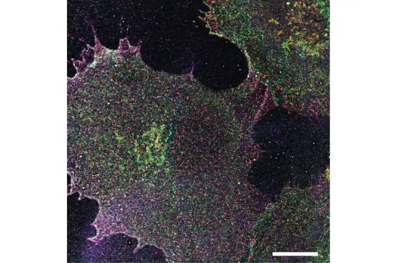
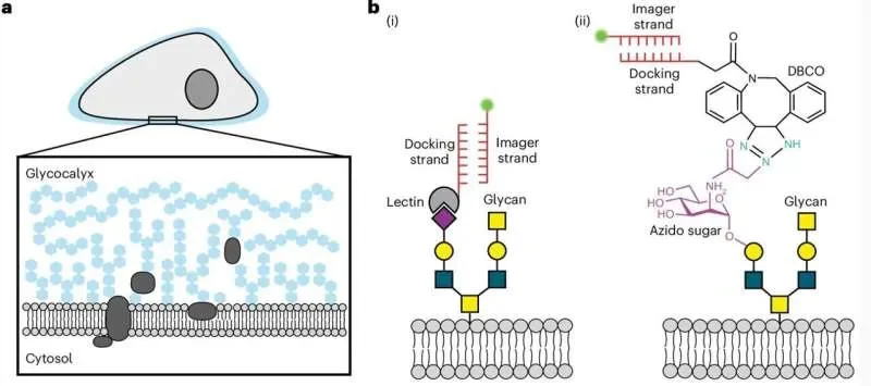
Researchers have utilized advanced super-resolution microscopy to visualize individual sugar structures on various cell types. This includes standard cell cultures, human blood cells, and tissue samples. The technique, dubbed 'Glycan Atlasing', provides the first direct observation of the glycocalyx acting as a display, broadcasting information about the cell's condition.
Mapping Internal States Externally
The core of this breakthrough lies in the Glycan Atlasing method. By examining the precise locations of complex carbohydrates on the cell's outer layer, scientists can now differentiate between various cellular conditions.
Read More: New Study Shows How Bacteria Make Energy, Could Lead to New Medicines
Early studies confirmed that the spatial distribution of these sugar structures can be reliably linked to distinct cell states.
This detailed mapping offers a new window into cell physiology, showing the glycocalyx as more than just a protective layer, but as an active information conduit.
Implications for Health and Disease
The ability to "read" cell surface sugars has profound implications for diagnostics and understanding disease progression.
Distinct glycan spatial patterns were observed in stimulated immune cells, mimicking an activated immune response, compared to their resting counterparts. This suggests potential for monitoring immune function with greater precision.
The research indicates that these surface patterns could also be used to identify different stages of cancer, potentially leading to earlier and more accurate detection.
The findings lay groundwork for advancements in personalized medicine, where an individual's cellular state could be assessed through their glycocalyx profile.
The Glycocalyx: A Cellular Information Hub
The glycocalyx, a complex mosaic of carbohydrates projecting from the cell membrane, is the cell's primary interface with its environment. It plays a crucial role in:
Mediating cellular signaling.
Facilitating immune recognition.
Organizing tissue structures.
This new method offers a deeper understanding of the glycocalyx's dynamic role in these fundamental biological processes. The research builds upon existing knowledge of cell surface structures and their interactions with the extracellular matrix, but elevates it to a new level of functional interpretation.