A novel nanoscopy method, developed at The Australian National University (ANU), has illuminated previously unseen communication pathways between living cells. This advanced technique allows researchers to observe cell interactions in three dimensions over extended periods, revealing dynamic, thread-like structures that transfer biochemical messages. The breakthrough, detailed in 'Nature Communications', promises to deepen our grasp of various human ailments and biological processes, including viral transmission.

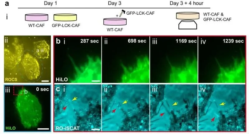
The core innovation lies in a 'label-free' nanoscopy approach called RO-iSCAT, which overcomes the limitations of traditional microscopes by providing high-resolution, three-dimensional views of cellular behaviors without requiring invasive dyes that can harm the cells. This enables scientists to track the formation, extension, retraction, and reconnection of nanoscale extensions over several days, uncovering intricate communication networks.

Unveiling Hidden Connections
For decades, the physical infrastructure of cellular communication has been a subject of speculation, largely due to technological constraints. Conventional microscopes struggled to capture the fine details of these transient structures.
Read More: New Hologram System Uses GPU to Measure Particles Faster

The ANU team's nanoscopy technique provides a crucial window into these elusive, thread-like cellular extensions.
These structures are vital for nearly all cellular signaling, communication, and movement.
The continuous imaging over days revealed highly organized networks facilitating the transfer of biochemical signals between neighboring cells.
Potential Impact on Disease and Viral Spread
The implications of this discovery extend to understanding and potentially treating diseases. The ability to visualize these cellular networks in action offers new avenues for research.

The technique has already been employed to study how human blood vessel cells connect to form new blood cells.
Crucially, it could shed light on how viruses spread, as many are believed to exploit these cellular bridges for transmission throughout the body.
A Milestone in Cellular Imaging
Researchers involved have described the ability to witness these slender tubes forming connections in real time as a significant achievement. The technology's capacity to observe dynamic motion, with structures twisting and forming stable bridges, marks a substantial leap in our observational capabilities. The research, while recent, has already garnered attention across multiple scientific news outlets, highlighting its perceived importance within the field.