Unveiling the Unseen Spin Dance
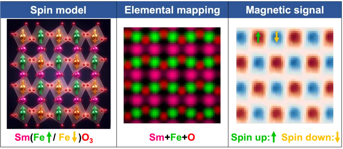
A novel imaging technique has peeled back layers of complexity in antiferromagnetic materials, exposing their hidden atomic-scale magnetic structures. This method, using electron magnetic circular dichroism (EMCD) with atomic-column resolution, marks a significant leap in characterizing materials previously opaque to detailed magnetic analysis. Researchers successfully demonstrated the technique on two types of antiferromagnets, DyFeO₃ and α-Fe₂O₃, revealing their intricate spin arrangements.
The significance lies in the potential of antiferromagnets for next-generation technologies. Their inherent properties— antiparallel atomic spins leading to zero net magnetization—render them exceptionally fast and resistant to external magnetic disturbances. These attributes make them prime candidates for developing high-speed, high-density 'spintronic' devices, which harness electron spin rather than just charge.
The Interface Enigma
One of the key findings involved an investigation at the interface between DyScO₃ and SmFeO₃. Here, the atomic-column EMCD technique precisely identified a 'magnetic dead layer'—a region with suppressed magnetic order—spanning just a single unit cell. This granular observation offers critical insights into how magnetic behavior is affected at material boundaries, a crucial factor in designing functional spintronic components. Such findings are vital for understanding and manipulating interfacial magnetic coupling, directly influencing interface engineering efforts for spintronic applications.
Read More: Heated Rocks Capture CO2 1000s of Times Faster
Technical Prowess
The breakthrough centers on an advanced application of EMCD, a method sensitive to the magnetic state of materials. By achieving resolution at the atomic column level, scientists can now map magnetic properties with unprecedented detail. This technique has overcome limitations that have long hindered the precise characterization of microscopic magnetic structures, especially within complex or buried interfaces. The experimental work also involved meticulous data processing procedures to extract these fine-grained EMCD signals, alongside performing magnetic measurements within an atomic-resolution electron microscope designed to operate in a field-free environment.
This development, published recently in Nature Nanotechnology, provides a powerful new lens through which to view and understand the subtle world of antiferromagnetic order, potentially accelerating the development of advanced electronic technologies.